Researchers find out how some sensory information breaks through to conscious experience.
11:00 AM
Author |
During our waking hours, the brain is receiving a near-constant influx of sensory signals of various strengths.
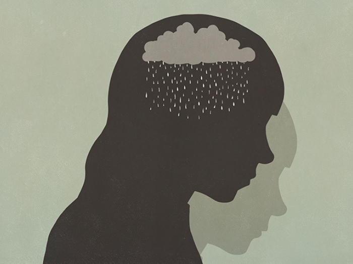
For decades, scientists have wondered why some signals rise to the light of conscious awareness while other signals of a similar strength remain in the dark shadows of unconsciousness. What controls the gate that separates the shadows and the light?
In a new study from the Department of Anesthesiology and Center for Consciousness Science at Michigan Medicine, researchers identify a key area in the cortex that appears to be the gate of conscious awareness.
"Information processing in the brain has two dimensions: sensory processing of the environment without awareness and the type that occurs when a stimulus reaches a certain level of importance and enters conscious awareness," explains Zirui Huang, Ph.D., research investigator in the Department of Anesthesiology.
Huang, along with lead researcher Anthony Hudetz, Ph.D. and their team, attempted to confirm that this switch occurs in a part of the brain called the anterior insular cortex, acting as a type of gate between low level sensory information and higher level awareness.
For the experiments, participants were put inside of a fMRI machine and administered the anesthetic drug propofol to control their level of consciousness. They were then asked to imagine themselves playing tennis, walking down a path or squeezing their hand, as well as asked to perform a motor activity (squeeze a rubber ball) as they gradually lost consciousness and regained it again after the propofol was stopped.
Previous research has shown that mental imagery produces brain activity similar to actually performing the activity. When participants imagine themselves playing tennis, the part of the brain responsible for controlling movement lights up. Other regions of the brain become deactivated when performing tasks, as mental attention is focused on the activity.
As the study participants began to lose consciousness, deactivations happened less frequently. When they completely lost consciousness, their corresponding brain areas also showed no activation in response to mental imagery tasks. As they regained some consciousness, they regained some activity related to mental imagery and with full consciousness shortly thereafter, their brain showed normal activation patterns.
MORE FROM THE LAB: Subscribe to our weekly newsletter
Looking for the correlation across these states of consciousness revealed activation of the anterior insular cortex played a role in the successful switch between these activations and deactivations.
"A sensory stimulus will normally activate the anterior insular cortex," says Hudetz. "But when you lose consciousness, the anterior insular cortex is deactivated and network shifts in the brain that support consciousness are disrupted." The anterior insular cortex, he explains, might act as a filter that allows only the most important information to enter conscious awareness.
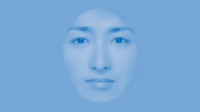
They sought to confirm their hypothesis with another classic psychological experiment, wherein a face is briefly flashed on a screen for a barely perceptible three hundredths of a second.
The face image is followed by a noisy high contrast image designed to interrupt conscious processing of the face image. Participants were then asked whether they saw a face or not. Whether the face was consciously accessed was predicted by activation in the anterior insular cortex.
"Anterior insular cortex has continuously fluctuating activity," says Huang. "Whether you can detect a stimulus depends upon the state of the anterior insula when the information arrives in your brain: if the insula's activity is high at the point of stimulus, you will see the image. Based on evidence from these two experiments, we conclude that the anterior insular cortex could be a gate for conscious awareness."
Paper cited: "Anterior insula regulates brain network transitions that gate conscious access," Cell Reports. DOI: 10.1016/j.celrep.2021.109081
Like Podcasts? Add the Michigan Medicine News Break on iTunes or anywhere you listen to podcasts.

Explore a variety of healthcare news & stories by visiting the Health Lab home page for more articles.

Department of Communication at Michigan Medicine
Want top health & research news weekly? Sign up for Health Lab’s newsletters today!