In recent years, the use of imaging tests has skyrocketed for post-treatment surveillance of thyroid cancer. But this hasn’t affected survival rates, new research shows.
6:30 PM
Author |
More imaging after thyroid cancer treatment identifies recurrence, but it does not always improve survival, a new study suggests.
SEE ALSO: International Collaboration Leads to Clues About Rare Cancer
Researchers from the University of Michigan Rogel Cancer Center looked at 28,220 patients diagnosed with differentiated thyroid cancer, using the Surveillance, Epidemiology and End Results-Medicare linked database.
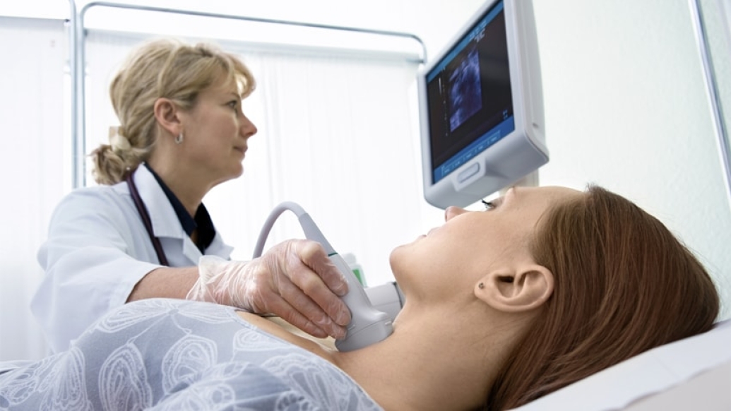
They used insurance claims data to track ultrasounds, PET scans and radioiodine scans in patients diagnosed with thyroid cancer from 1998 to 2011. These scans would be done to monitor for signs of cancer returning.
Researchers found 57 percent of patients had at least one ultrasound, 24 percent had a radioiodine scan and 15 percent had a PET scan. Patients who had these tests were more likely to have additional treatments, such as surgery, radioactive iodine treatment or radiation therapy.
Still, although the use of imaging rose substantially during this time, the overall death rate from the cancer did not change, according to the study published in BMJ.
"Over time, we have seen this marked increase in the use of imaging after primary treatment of thyroid cancer despite the fact that the majority of our patients have low-risk cancer. For the most part, this imaging isn't affecting survival," says study author Megan R. Haymart, M.D., assistant professor of medicine at the University of Michigan Medical School.
"With this post-treatment surveillance imaging, we're picking up more recurrences. But is that clinically significant? We might be picking up really small lymph nodes that, if left untreated, wouldn't have impacted survival," says study author Mousumi Banerjee, Ph.D., research professor of biostatistics at the University of Michigan School of Public Health.
In the study, only radioiodine scans led to improved survival, the researchers found. Ideally, these scans are used when blood tests suggest an increase in a certain tumor marker and the patient is known to respond to radioactive iodine treatment.
Just because we can image doesn't mean we should for all. We need to consider whether it is the appropriate thing to do.Mousumi Banerjee, Ph.D.
When is post-treatment imaging appropriate?
More people are being diagnosed with low-risk thyroid cancer, but the use of imaging among these patients has skyrocketed disproportionately. Thyroid cancer generally has a high survival rate — about 96 percent of patients are alive 10 years later. But a small number of thyroid cancers are more aggressive and likely to return.
SEE ALSO: When It's Not Exactly Cancer: Renaming Early Stage Thyroid Tumors
"There is a place for imaging in thyroid cancer survivors. But the specific type of imaging needs to be tailored to the patient," Haymart says. "When we have a patient with a favorable prognosis, certain types of imaging may not be necessary. But there is a group for whom it might be appropriate."
Researchers have raised the question of what kind of surveillance regimen is appropriate after treatment for many types of cancer, including lung cancer and breast cancer. The Choosing Wisely campaign aims to create a national dialogue about avoiding unnecessary medical tests. This study highlights the importance of reassessing appropriate tests after cancer treatment.
It's an important question because imaging is not without impact, say Haymart and Banerjee, both members of the University of Michigan Institute for Healthcare Policy and Innovation.
Although potential physical harm from these tests is low, many cancer patients report "scanxiety," feelings of intense distress before imaging as they fear bad news. The tests can also be costly. All of this is compounded if the tests lead to additional treatment, with some of these treatments having downstream risks.
"The impact of these tests on patients' psycho-social well-being is also important. Just because we can image doesn't mean we should for all. We need to consider whether it is the appropriate thing to do. There is a large group of patients for whom some of these imaging tests may be unnecessary," Banerjee says.
Next steps for this work include examining the tests' cost-effectiveness, performing randomized controlled trials of surveillance imaging, and understanding whether patients or providers are pushing for these tests.

Explore a variety of healthcare news & stories by visiting the Health Lab home page for more articles.

Department of Communication at Michigan Medicine
Want top health & research news weekly? Sign up for Health Lab’s newsletters today!