For a long time, nonepileptic seizures were not believed to involve structural changes in the brain.
5:00 AM
Author |
There are just over 3 million Americans with epilepsy who experience seizures due to abnormal electrical activity in the brain.
A smaller group of people also have seizures not caused by epilepsy – known by many names, including functional seizures, psychogenic seizures, nonepileptic seizures, or even the pejorative term pseudoseizures. Scientists have long understood these as the body's response to mental stressors, like anxiety and post-traumatic stress disorder. But a new study finds that functional seizures are associated with structural changes in the brain that can be seen using MRI.
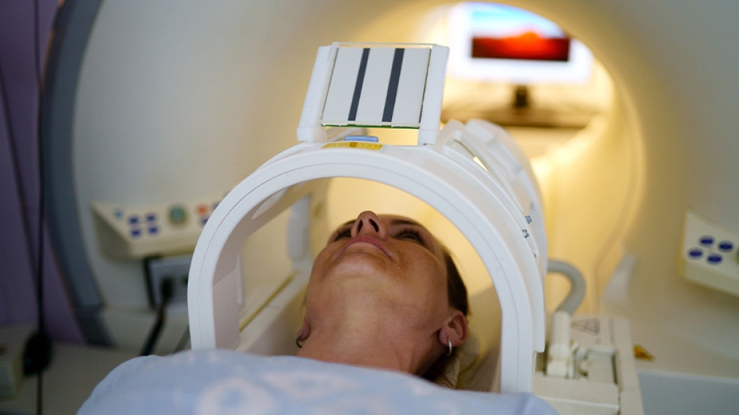
The team of researchers led by Michigan Medicine analyzed more than 650 clinical-grade MRIs, comparing images from patients with functional seizures to those who did not experience seizures and could have other mental health conditions, such as depression and anxiety.
SEE ALSO: Sleep problems pervasive and persistent among children with epilepsy (uofmhealth.org)
Results published in Epilepsy & Behavior reveal that patients with functional seizures had thinning in the superior temporal cortex, which affects a person's cognitive awareness and control of one's actions, and thickness of the left occipital cortex, responsible for the processing of visual and other sensory information. These changes were not present in people who had depression, anxiety and obsessive compulsive disorder (OCD).
"If we can figure out what brain changes make people have functional seizures, we can begin to look at how we can change that back," said first author Wesley Kerr, M.D., Ph.D., a neurologist and epileptologist at University of Michigan Health.
Like Podcasts? Add the Michigan Medicine News Break on Spotify, Apple Podcasts or anywhere you listen to podcasts.
"Right now, the treatment for functional seizures is cognitive behavioral-informed therapy, but this therapy isn't effective for everyone. We hope to find new ways to target treatments to help patients have fewer seizures and better quality of life."
Epilepsy misdiagnosis
One of the challenges in treating functional seizures is identifying them early enough. Most patients with functional seizures are treated for epilepsy for an average of nine years before being correctly diagnosed.
That misdiagnosis can cause real harm to patients, says Kerr, a clinical instructor of neurology at U-M Medical school. In the future, this type of structural MRI may reveal biomarkers that differentiate the two conditions.
"During the years before people are accurately diagnosed with functional seizures, 80% of them were prescribed anti-seizure medications used to treat epilepsy," he said. "Those medications have side-effects, including fatigue and dizziness. These people can also come into the ER with prolonged seizures and end up being intubated and given high doses of medication, which can be lifesaving for someone with epilepsy but is both not needed and dangerous for functional seizures."
If we can figure out what brain changes make people have functional seizures, we can begin to look at how we can change that back.Wesley Kerr, M.D., Ph.D.
Psychotherapy is used as a treatment for functional seizures to address the underlying psychological stressors that are overflowing and leading to symptoms.
Differentiating between epileptic and nonepileptic seizures
While epilepsy has the distinct marker of electrical abnormality, discerning between the condition and a functional seizure is not simple. Clinicians diagnose epilepsy based on the number and type of seizures someone experiences, along with diagnostic testing. If seizures are epileptic, not acting quickly could cause permanent brain damage or death.
"In those cases, people tend to give high doses of seizure medication if they are not sure," Kerr said. "And around 10% of people who come in with what we think is status epilepticus are actually having prolonged functional seizures."
SEE ALSO: Cost of brand-name epilepsy drugs increased by 277% over 8 years (uofmhealth.org)
After hearing about or observing a seizure, providers can take an electroencephalogram, or EEG, which picks up electrical signals in the brain. However, about half of people with known focal epilepsy have EEGs that are read as normal.
"We would always like to have the EEG and MRI information to back up what we see in the patient clinically, but we still need to see the seizure to diagnose functional seizures reliably," Kerr said.
"But once we diagnose someone with functional seizures, we can reduce the high health care utilization they have from coming into the ER often and having to pay a lot for anti-seizure medications to improve their quality of life. We do better when we can diagnose people with functional seizures earlier."
As researchers continue to explore the structural differences in changes to the brain between functional seizures and epilepsy, MRI will continue playing a key role in improving the diagnosis and treatment of both conditions.
"We know that early, accurate diagnosis of functional seizures is essential to starting treatment that will improve quality of life," said co-author Nicholas J. Beimer, M.D., clinical assistant professor of neurology and psychiatry at U-M Medical School.
"This work is an example of how we can look more carefully at the tests we already perform, like a standard MRI of the brain, for subtle clues that lead us to the correct diagnosis. I look forward to how this research will be used to improve our diagnostic speed and accuracy, so that we can get our patients with functional seizures to the right treatment sooner."
Live your healthiest life: Get tips from top experts weekly. Subscribe to the Michigan Health blog newsletter
Headlines from the frontlines: The power of scientific discovery harnessed and delivered to your inbox every week. Subscribe to the Michigan Health Lab blog newsletter
Additional authors include Lubomir M. Hadjiski, Ph.D., William C. Stacey, M.D., Ph.D., both of Michigan Medicine, John M. Stern, M.D., Hiroyuki Tatekawa, M.D., John K. Lee, M.D., Ph.D., Amir H. Karimi, Siddhika S Sreenivasan, Jena M. Smith, L. Brian Hickman, M.D., Jerome Engel Jr., M.D., Ph.D., Noriko Salamon, M.D., Dawn S. Eliashiv, M.D., all of David Geffen School of Medicine at UCLA, Joseph O'Neill, Ph.D., Jane & Terry Semel Institute for Neuroscience and Human Behavior, Ivanka Savic, M.D., Ph.D., Nilab Nasrullah, both of Karolinska University Hospital, Randall Espinoza, M.D., Katherine Narr, Ph.D., Jamie D. Feusner, M.D., all of University of California Los Angeles.
Engel, Stern, and Kerr have received speaking fees and honoraria for articles on this topic.
Paper cited: "Clinical MRI morphological analysis of functional seizures compared to seizure-naïve and psychiatric controls," Epilepsy & Behavior. DOI: 0.1016/j.yebeh.2022.108858

Explore a variety of healthcare news & stories by visiting the Health Lab home page for more articles.
Department of Communication at Michigan Medicine
Want top health & research news weekly? Sign up for Health Lab’s newsletters today!