For surgeons, the SRS microscope offers speed, safety and efficiency in assessing what is and is not tumor tissue — on the spot.
4:34 PM
Author |
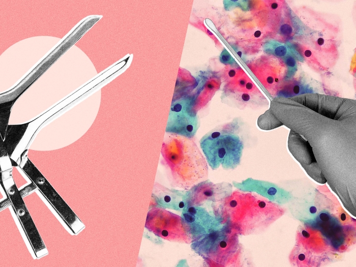
Where does a brain tumor start and healthy brain tissue begin? This is one of the most challenging questions a brain surgeon faces, because brain tumor tissue can be difficult to distinguish from the rest of the brain.
That's one of many reasons we are testing a new microscope designed to radically change brain tumor surgery, with an eye on both patient safety and better efficiency. So far, we have used the microscope — called the stimulated Raman scattering (SRS) microscope — on tissues from 89 patients with great success.
Two critical factors: timing and location
On top of the challenge of visually locating the edges of a tumor, we also typically need to wait 30–45 minutes for a frozen tumor section to be created and interpreted using traditional techniques.
Not so with the SRS microscope. It allows us to detect a tumor in a few seconds.
Right now, we are using the microscope on an experimental basis through grants from the National Institutes of Health and the University of Michigan Translational Research and Commercialization for Life Sciences Program. We are using the microscope almost exclusively on neurosurgical cases. I'm also collaborating with Matt Spector, M.D., who is a head and neck surgeon, to look at squamous cell carcinoma.
Our work suggests that surgeons also will be able to use the SRS microscope in breast cancer surgery and for other cancerous tumors. Ultimately, the goal is to use a data-driven, objective approach to determine the exact location of a tumor infiltration instead of relying exclusively on a surgeon's intuition.
The SRS microscope can tell you on the spot, in the operating room, whether tissue contains a tumor or not.Daniel Orringer, M.D.
How we achieve speed
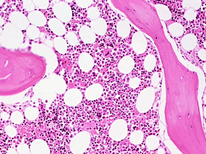
The standard pathology lab workflow used for 150 years requires removing the tumor tissue, then fixing, embedding, staining with dyes and mounting the tissue on slides.
With the SRS technology, we simply load the tissue onto the SRS microscope and take an image of it. With an SRS microscope, images are created based only on the chemical building blocks that exist in the tissue. There are no other steps — no dyes, no fixation and no sectioning. The images we get are comparable to the images we get with traditional microscopes. The SRS microscope can tell you on the spot, in the operating room, whether tissue contains a tumor or not.
The SRS technology we are using was originally developed in the laboratory of my collaborator at Harvard, Professor Sunney Xie, and built by a company called Invenio Imaging Inc.
What comes next
We are also working on a second-generation SRS microscope that will sit close to the operating table. The goal is for the entire medical team to be able to use and understand the device easily so the images will help determine immediately whether the surgeon needs to remove more tissue.
We hope to have the machine approved by the FDA within 18 to 24 months.
For surgeons, we want the SRS to provide precision, efficiency and speed. Ultimately, that means better surgical outcomes for patients.
Disclosure: Orringer and Xie are advisers and shareholders of Invenio Imaging Inc., a company developing SRS microscopy systems. Their co-author Freudiger is an employee of Invenio Imaging, Inc.
The University of Michigan, Harvard University and Invenio Imaging have intellectual property used in this research.

Explore a variety of healthcare news & stories by visiting the Health Lab home page for more articles.
Department of Communication at Michigan Medicine
Want top health & research news weekly? Sign up for Health Lab’s newsletters today!