A complex camera has the potential to help doctors get a clearer, more accurate look at the human eye. How a trio of U-M experts from different fields made the discovery.
1:00 PM
Author |
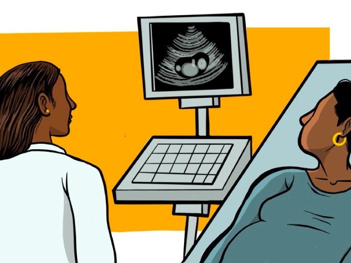
In a typical examination, ophthalmologists explore a patient's eye depths using light and magnification, and also take pictures to look for problems and monitor changes over time.
MORE FROM THE LAB: Subscribe to our weekly newsletter
Clinicians must be intuitive, using their own sight and training to assess whether any abnormalities are present in those images.
But these exams, a critical first step in preventing or treating blindness and other serious conditions, aren't available to many people who need them.
And existing methods of three-dimensional eye imaging can have limitations.
A new 3-D photographic technique, adapted from automotive engineering, could offer a fast and simple way to capture the eye's intricate features with richer detail — and might one day be used to help people obtain access to specialty eye care remotely.
The means: a plenoptic camera that can essentially take thousands of photos of the same object at a single moment in time from slightly different directions, heights and distances.
"By using this camera, we can create a permanent digital record of the same kind of images that ophthalmologists form in their minds through physical examinations, which can be examined at any time, and examined over time to measure changes," says Maria Woodward, M.D., M.S., an ophthalmologist at the University of Michigan Kellogg Eye Center.
The work, part of a multidisciplinary research initiative known as MCubed, marks a unique collaboration between U-M ophthalmologists and systems engineers.
Woodward, who specializes in conditions affecting the front part of the eye, was looking for new imaging modalities that could better capture structures in this area, such as the iris and lens. She teamed up with David Burke, Ph.D., a professor of human genetics, and Volker Sick, a professor of mechanical engineering, for the effort.
It was Sick who recognized that the plenoptic technology he uses to study fluid dynamics in optical engine research could be applied to create enhanced imaging of the human iris.
That hunch has brought added clarity to Woodward's cause.
Improved imaging technology
Right now, 3-D eye imaging in ophthalmology is made possible by combining a sequence of two-dimensional cross-sectional images into a single picture.
SEE ALSO: Telemedicine Could Improve Eye Exam Access for People with Diabetes
The physician's expertise lies in his or her ability to form a 3-D image of the patient's eyes, to compare that image to her knowledge of what "normal" or "healthy" eyes are supposed to look like, and to monitor the patient for changes over multiple visits.
But one problem with this approach is that a patient's eye structures can morph during the course of recording these images due to the dynamic nature of eye tissue as well as the patient's natural eye movements. Such imaging also requires the eye to be anesthetized because it must come in contact with the camera probe.
In addition, these images must be taken by a highly specialized photographer due to the difficulty of the technique.
A plenoptic camera can improve on these techniques by capturing the intensity of the light source as well as the angle of each ray. These photos are then reconstructed into a single enhanced "4-D" image that's richer and more accurate than what is possible through normal human vision.
Furthermore, the plenoptic camera doesn't need to make physical contact with the eye, and it is relatively easy to use.
The benefits may have big implications: In their research, the MCubed team demonstrated that a plenoptic camera system could be used to capture 3-D iris patterns in a single photograph with enough accuracy and precision to make disease diagnosis and continued monitoring feasible.
They recently published their findings in Biomedical Optics Express.
Looking forward, in the same way that we can learn about eye health when we examine the eye, we can also learn about a patient's blood vessels and nerves.Maria Woodward, M.D., M.S.
Potential to broaden reach
At the moment, image-based telemedicine within ophthalmology is primarily used to monitor patients over time for sight-threatening diseases, such as retinopathy in premature infants and people with diabetes.
But Woodward says there is great interest in moving beyond these applications to extend specialty eye care to different settings such as emergency rooms (since about 2 million people each year in the U.S. head to the emergency department for eye problems rather than an ophthalmologist).
Because lower-cost versions of the plenoptic camera exist, it also has potential to extend to rural and urban areas, where access to this care is more limited. A remote person, then, might receive the camera to take and submit images before returning the equipment.
"Physicians could 'see' patients to know who is sick, regardless of their geographical location," Woodward, who is also a member of the U-M Institute for Healthcare Policy & Innovation, says. She notes that the technology would have to be carefully implemented to allow doctors to also treat patients, when necessary.
Other fields of medicine that are actively using or exploring image-based telemedicine offer some very translatable examples of modalities that could be extended into ophthalmology, such as radiology, dermatology and mental health, Woodward says.
Reimbursement has been one barrier to expanding telemedicine applications. "Payment shouldn't happen unless a modality is safe, effective and valuable," Woodward says. "We need to continue to innovate in this area to show we can reach more people and still maintain high-quality care."
Next steps
To move this technology forward, the team hopes to optimize the plenoptic camera's capabilities for use in eye exams and also test its capabilities for examining other structures of the eye beyond the iris.
They would also like to investigate whether less expensive, more portable plenoptic camera models could produce images of the same or similar quality, resulting in wide clinical utility and broader applications beyond eye health.
"Looking forward, in the same way that we can learn about eye health when we examine the eye, we can also learn about a patient's blood vessels and nerves," Woodward says. "We could potentially use this tool to study diabetes, brain injury or vascular diseases."
Explore a variety of healthcare news & stories by visiting the Health Lab home page for more articles.
Department of Communication at Michigan Medicine
Want top health & research news weekly? Sign up for Health Lab’s newsletters today!