Researchers track protein binding, build synthetic proteins to study gene expression.
5:00 AM
Author |
How does a nose remember that it's a nose? Or an eye remember that it's an eye?
As scientists probe the question of how cells remember what kind of cells they are supposed to be, or their genetic lineage, it's important to understand how cells express different genes without changing the DNA sequence itself.
But studying this subject is difficult: Researchers can purify the proteins that drive genetic expression, put them in a test tube and watch them bind. But doing so inside the nucleus of cells, their native environment, has been so far impossible.
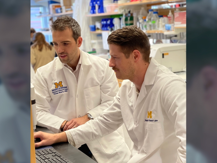
Now, a team of researchers at three University of Michigan labs have been able to track how a protein binds to its chromatin substrate within a living cell by establishing a collaboration that combines state-of-the-art ultra high-resolution imaging, synthetic protein design and computational modeling. Their results are published in Science Advances.
"The biological question that we're asking is, 'How do cells actually remember past experiences? And how do these experiences also lead to cells establishing distinct identities, as it happens in the case of the human body where you have lineages of cells that form neurons, or blood cells, or brain cells, and all actually maintain their identities for many generations,'" said lead author Kaushik Ragunathan, Ph.D., assistant professor of biological chemistry at the U-M Medical School.
"An example I like to think about is that if you chop off your nose, you don't get a hand growing there, even though the genome in your nose and the genome in your hand are exactly the same."
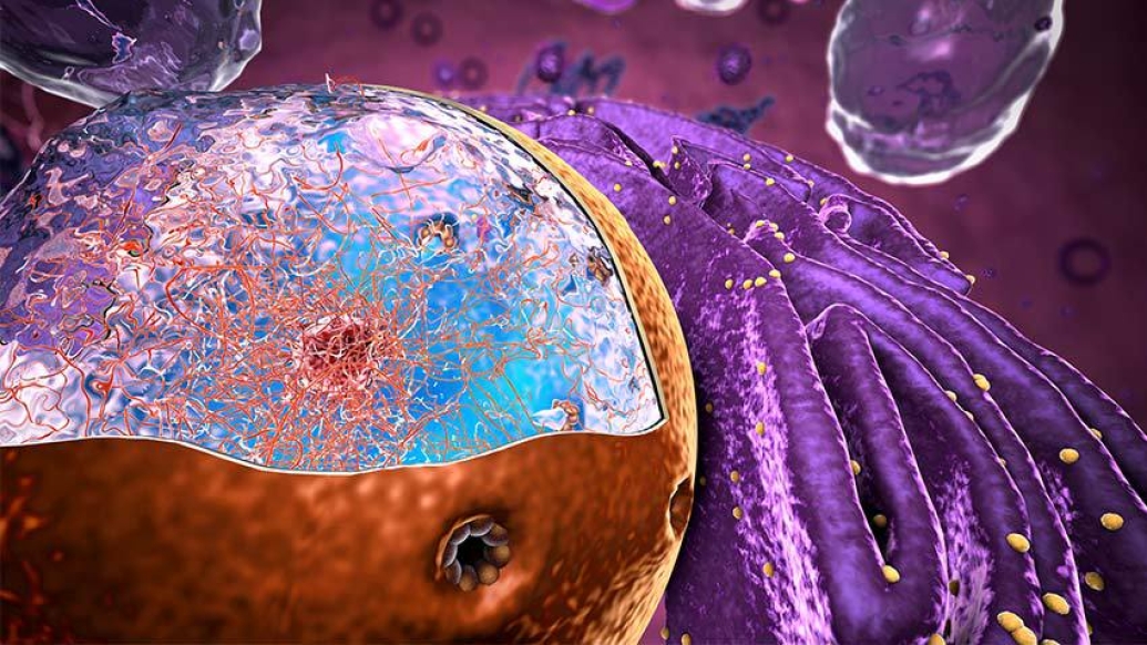
Cells control how and which genes are expressed from a copy of the DNA sequence held within each cell, despite that sequence being the same across all cells in the body. One way they control expression is by changing how tightly the DNA is packaged within the nucleus using proteins called "histones." Histones can be modified through the addition of small chemical tags that regulate how tightly the DNA is wound around them and thus whether the genes can be expressed.
Proteins that have the ability to read, write and erase these histone tags explore the DNA within the nucleus of the cell very rapidly—on the order of milliseconds, according to Ragunathan. Ultimately, all this epigenetic information needs to be inherited across generations, but the recognition of these tags is a complicated process that involves chromatin binding and proteins meeting and interacting with each other amidst the chaos of all other possible competing interactions within the cell.
Being able to understand each step of the process—and therefore enabling control of how the epigenetic information is inherited—intrigued co-author Julie Biteen Ph.D., professor of chemistry and biophysics.
Biteen uses single-molecule fluorescence imaging to track individual proteins inside cells. Her lab can see where these proteins are relative to the chromatin, and Ragunathan's expertise is in the molecular mechanisms underpinning how histone modifications and histone-binding proteins interact. These two worlds needed to come together so that the biochemistry of what happens in a test tube outside of cells could be tested to figure out what happens inside of them.
"The timing of this process is critically important to ensure that the right genes are silenced at the right place and at the right time," Biteen said. "What hooked me on this project is that in vitro—in a test tube—you can purify two proteins, watch them bind and see how good that binding is, or what is the affinity for one another. That tells you what can happen in the cells, but not what does happen in the cells."
Biteen and Ragunathan worked with Peter Freddolino, Ph.D., associate professor of biological chemistry, and computational medicine and bioinformatics at the U-M Medical School, to combine computer modeling with their experimental results.
"This is really where our collaboration becomes really powerful," Biteen said. "On one hand, seeing molecules is very helpful and knowing how fast the molecules move helps a lot in terms of understanding what is possible inside the cell, but here we could take a leap forward by perturbing the system even in unnatural ways in order to understand what these different motions of molecules in the cell actually mean."
While epigenetic marks are tremendously important for maintaining different tissues in complex organisms like humans, they also play an important role in regulating genes of single-celled organisms such as yeast. The team focused on a type of HP1 protein in yeast cells called Swi6. This family of proteins binds to a specific type of histone modifications in the cell to enforce gene silencing. By integrating fluorescent labels with Swi6, Bitee''s lab watched Swi6 move inside the cell's nucleus.
While Swi6 searches for the correct binding site on DNA, it moves quickly, Biteen said. When it finds its target, it slows down significantly. The movement of a protein within the cell is akin to gears in a car and things can move at different speeds based on whom proteins interact with.
"From these spaghetti tracks that we get inside the cell, we then figure out how much time they are spending searching and how much of the time they are spending bound," Biteen said. "The amount of time they spend not moving tells us about how strongly they're interacting and their biochemical properties."
While Biteen's lab can measure movements in the cell on the scale of tens of milliseconds, much of the biochemistry happening in the cell is happening even faster, she said. Freddolino took this experimental information and developed models to estimate the ability of the Swi6 proteins to jump between the binding states that were identified in experiments.
Freddolino's modeling took into account the experimental measurements and the possible biochemical properties, which includes how the Swi6 molecules interact in the cell. These interactions include molecules that freely float in the solution of the cell, molecules that have bound to DNA, and molecules that are "holding hands" with each other, he said.
"My lab wanted to come up with a more fine-grained model that estimated what was the most likely set of molecular states of the proteins and their ability to jump between those states, that would then give rise to the imaging data that Biteen's lab created," Freddolino said.
"Having this numerical model allows us to do the computational experiments of what happens if the protein binding is twice as fast as we think. What if it's 10 times as fast as we think? Or 10 times slower? Could that still give rise to the data? Very happily, in this case, we were able to show that the relevant processes were really being captured in the fluorescence microscopy."
After identifying the binding properties of natural Swi6, the researchers tested their findings by redesigning Swi6 from its components to see whether they could replicate some of its biochemical properties, Ragunathan said. This allowed the researchers to determine that the imaging and modeling conducted in the first part of the paper reflects how the protein was binding in its native environment.
"Can we do what nature did over the course of millions of years and make a protein that in many ways has properties similar to that of Swi6 in cells?" Ragunathan said. "In vivo biochemistry, which is what we've decided to call this, was not something that was ever thought to be possible inside living cells, but we have shown this is entirely feasible by using imaging as a modality. We are using this project as a foundation in order to understand how these epigenetic states can be established and maintained across generations."
Live your healthiest life: Get tips from top experts weekly. Subscribe to the Michigan Health blog newsletter
Headlines from the frontlines: The power of scientific discovery harnessed and delivered to your inbox every week. Subscribe to the Michigan Health Lab blog newsletter
Like Podcasts? Add the Michigan Medicine News Break on Spotify, Apple Podcasts or anywhere you listen to podcasts.

Explore a variety of healthcare news & stories by visiting the Health Lab home page for more articles.
Department of Communication at Michigan Medicine
Want top health & research news weekly? Sign up for Health Lab’s newsletters today!