The tiny, scaffoldlike device facilitates early detection of metastatic cancer cells and improves survival, a new study finds.
7:00 AM
Author |

A small device implanted under the skin can improve breast cancer survival by catching cancer cells, slowing the development of metastatic tumors in other organs and allowing time to intervene with surgery or other therapies.
MORE FROM THE LAB: Subscribe to our weekly newsletter
These findings, reported in Cancer Research, suggest a path for identifying metastatic cancer early and intervening to improve outcomes.
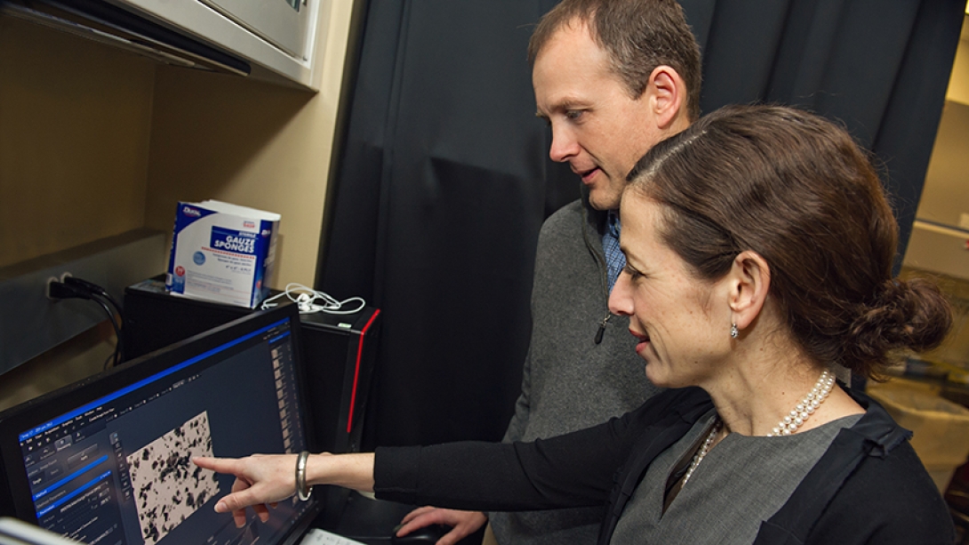
"This study shows that in the metastatic setting, early detection combined with a therapeutic intervention can improve outcomes. Early detection of a primary tumor is generally associated with improved outcomes. But that's not necessarily been tested in metastatic cancer," says study author Lonnie D. Shea, Ph.D., William and Valerie Hall Chair of Biomedical Engineering at the University of Michigan.
The study, done in mice, expands on earlier research from this team showing that the implantable scaffold device effectively captures metastatic cancer cells. Here, the researchers improve upon their device and show that surgery prior to the first signs of metastatic cancer improved survival.
"Currently, early signs of metastasis can be difficult to detect. Imaging may be done once a patient experiences symptoms, but that implies the burden of disease may already be substantial," says study author Jacqueline S. Jeruss, M.D., Ph.D., associate professor of surgery and biomedical engineering and director of the Breast Care Center at the University of Michigan Rogel Cancer Center.
"Improved detection methods are needed to identify metastasis at a point when targeted treatments can have a significant beneficial impact on slowing disease progression."
Our results suggest that bringing immune cells into the scaffold limits the ability of those immune cells to prepare the metastatic sites for the cancer cells.Lonnie D. Shea, Ph.D.
Setting a trap for cancer cells
The scaffold is made of microporous poly(ɛ-caprolactone) (PCL), a material commonly used in sutures and wound dressings and approved by the Food and Drug Administration. It's biodegradable and can last up to two years within a patient. The researchers envision it implanted under the skin, monitored with noninvasive imaging and removed upon signs of cancer cell colonization, at which point treatment could be administered.
SEE ALSO: For Breast Cancer, When Does Worry Outweigh Risk?
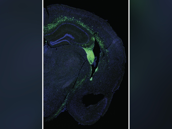
The scaffold is designed to mimic the environment in other organs before cancer cells migrate there. The scaffold attracts the body's immune cells, and the immune cells draw in the cancer cells. This then limits the immune cells from heading to the lungs, liver or brain, where breast cancer commonly spreads.
"Typically, immune cells initially colonize a metastatic site and then pave the way for cancer cells to spread to that organ. Our results suggest that bringing immune cells into the scaffold limits the ability of those immune cells to prepare the metastatic sites for the cancer cells. Having more immune cells in the scaffold attracts more cancer cells to this engineered environment," Shea says.
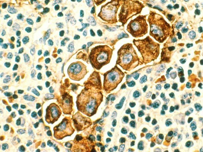
In the mouse study at five days after tumor initiation, researchers found a detectable percentage of tumor cells within the scaffold, but none in the lungs, liver or brain, suggesting that the cancer cells hit the scaffold first.
At 15 days after tumor initiation, they found 64 percent fewer cancer cells in the livers and 75 percent fewer cancer cells in the brains of mice with scaffolds compared with mice without scaffolds. This suggests that the presence of the scaffold slows the progress of metastatic disease.
The researchers removed the tumors at Day 10, which is after detection but before substantial spreading, and found the mice that had a scaffold in place survived longer than mice that did not have a scaffold. While surgery was the primary intervention in this study, the researchers suggest that additional medical treatments might also be tested as early interventions.
In the mouse study at five days after tumor initiation, researchers found a detectable percentage of tumor cells within the scaffold, but none in the lungs, liver or brain, suggesting that the cancer cells hit the scaffold first.
At 15 days after tumor initiation, they found 64 percent fewer cancer cells in the livers and 75 percent fewer cancer cells in the brains of mice with scaffolds compared with mice without scaffolds. This suggests that the presence of the scaffold slows the progress of metastatic disease.
SEE ALSO: Determining When Early Breast Cancer Is Aggressive — and When It's Not
The researchers removed the tumors at Day 10, which is after detection but before substantial spreading, and found the mice that had a scaffold in place survived longer than mice that did not have a scaffold. While surgery was the primary intervention in this study, the researchers suggest that additional medical treatments might also be tested as early interventions.
In addition, researchers hope that by removing the scaffold and examining the cancer cells within it, they can use precision medicine techniques to target the treatment most likely to have an impact.
This system is early detection and treatment, not a cure, the researchers emphasize. The scaffold won't prevent metastatic disease or reverse disease progression for patients with established metastatic cancer.
The team will develop a clinical trial protocol using the scaffold to monitor for metastasis in patients treated for early stage breast cancer. In time, the researchers hope it could also be used to monitor for breast cancer in people who are at high risk because of genetic susceptibility. Researchers are also testing the device in other types of cancer.

Explore a variety of healthcare news & stories by visiting the Health Lab home page for more articles.

Department of Communication at Michigan Medicine
Want top health & research news weekly? Sign up for Health Lab’s newsletters today!